
How to Get Rid of Milia Under the Eyes: Safe Methods
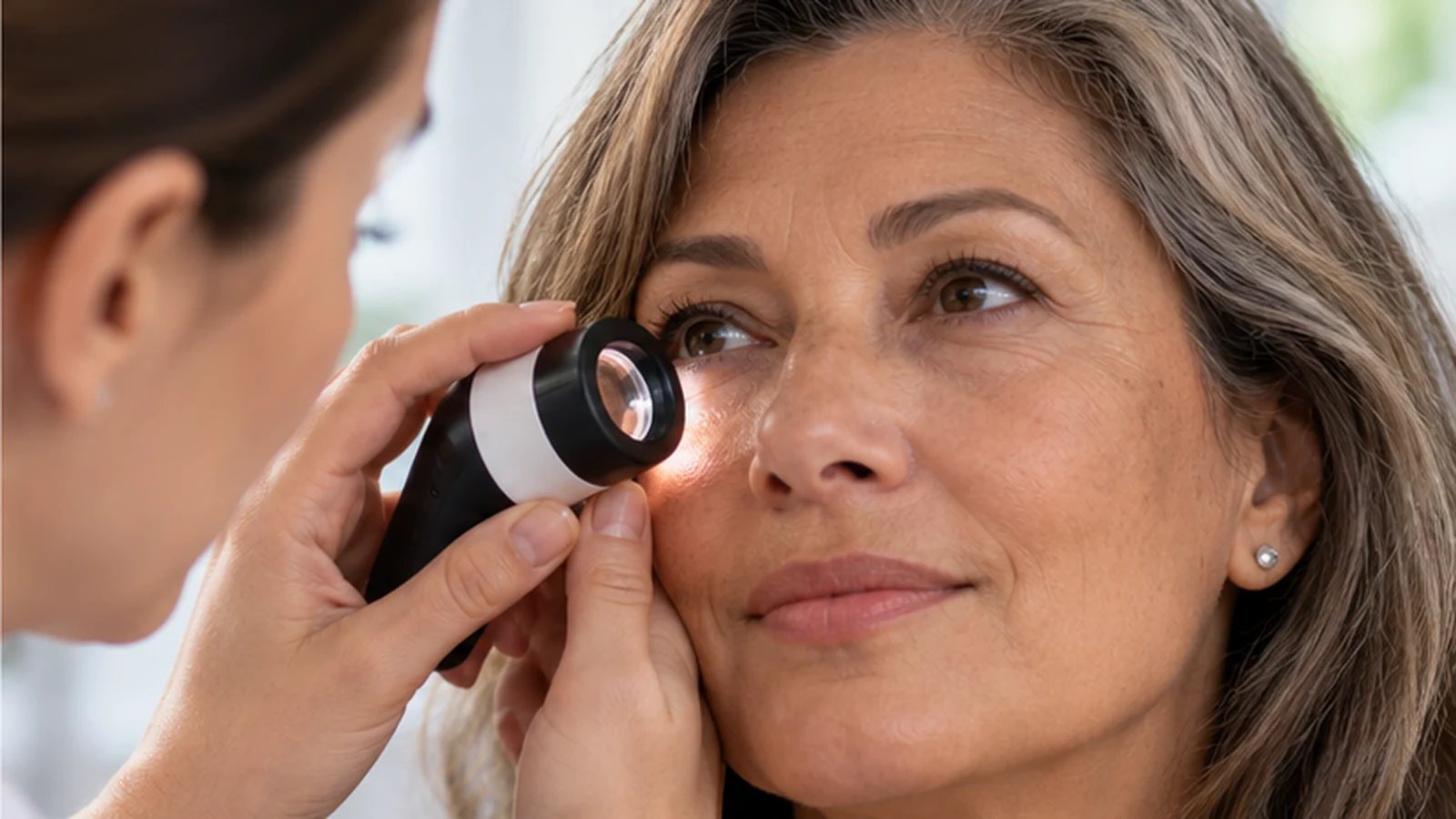
If you have noticed one or several firm white dots beneath your lower eyelid, you may already have tried to cover them with concealer. They do not hurt and they cannot be squeezed out like an ordinary spot. If these are milia, the safest way to remove them promptly is to have a dermatologist confirm the diagnosis and, if appropriate, open the tiny superficial cyst and extract its contents professionally. Do not pierce or squeeze a spot at home, especially near the lash line: the thin skin and the eye are vulnerable to injury. Small milia sometimes disappear without treatment, so appearance alone rarely makes removal urgent.
What can you do today without injuring the skin?
A milium is a small cyst containing keratin, a protein found in surface skin cells. It has no normal opening to the surface, so it is not a clogged pore that pressure can clear. Until you know the diagnosis, leave it alone. Cleanse gently, pat rather than rub the area dry and protect your face from sunlight. These steps care for the skin; they do not promise to dissolve an established cyst.
Avoid needles, lancets, tweezers, comedone extractors, alcohol, caustic herbal products, at-home freezing devices and cautery tools. DIY removal can cause a scar, pigment change, infection or an eye injury. Rough scrubs and strong acids are not made safe by applying them “just to the spot” next to an eyelid.
If you recently changed to a thick cream, balm or ointment, note its name and show the packaging to the clinician. Some milia occur after skin injury or in association with occlusive products, but one jar does not explain every case. Do not stop a prescribed medicated ointment, especially a steroid, on your own: discuss a change with its prescriber.
A similar white dot is not always a milium
Many different bumps look alike in online photographs. Under the eyes, syringomas (benign growths of sweat-duct tissue), xanthelasma (yellowish plaques), closed comedones, other cysts and, less often, lesions requiring further assessment can resemble milia. Similar colour does not mean identical treatment. Never choose a procedure from a photo alone, particularly if a lesion sits on the lid margin or is changing.
| What you notice | What it may mean | A sensible next step |
|---|---|---|
| One firm, painless white or pearly dot | Possibly a single milium, but description alone cannot establish the diagnosis | Leave it alone; discuss observation or office removal |
| Many small, skin-coloured bumps beneath both eyes | Could be syringomas or other lesions rather than dozens of milia | See a dermatologist first; treatment differs |
| A flat, yellowish plaque near the inner corner of the eye | Possible xanthelasma | Ask for diagnostic confirmation and, when appropriate, discuss lipid assessment |
| A bump that grows, bleeds, crusts or changes the eyelashes | Atypical for an uncomplicated, stable milium | Have it medically assessed before any cosmetic removal |
Marked redness, increasing pain, substantial swelling, pus, reduced vision or pain in the eye itself warrant medical attention rather than waiting for a cosmetic appointment. For lesions touching the ocular surface or lash margin, an ophthalmologist experienced in eyelid disorders may be needed.
How does a clinician remove a confirmed milium?
For a small solitary lesion, a clinician may carefully open the superficial roof using a sterile instrument and remove the keratin. This is different from forcefully squeezing an inflamed pimple. The technique depends on depth, position, number of bumps, skin condition and proximity to the eye. You may feel a brief prick or pressure; discomfort differs with the technique and the person. Ask whether local anaesthesia is needed and who will perform a procedure so close to the lid.
For numerous or persistent milia, published options include curettage (removal using a small scraping instrument), electrosurgery, sometimes freezing, laser treatment and topical retinoids. Listing a method does not make it suitable for delicate under-eye skin. The closer the lash margin and eye surface, the more important specialist experience and proper eye protection become. Laser is neither mandatory nor automatically gentler; burns, pigment changes and scarring are possible.
| Option | What it offers | What it cannot promise |
|---|---|---|
| Watch and wait | No procedure-related injury or expense | An adult milium may persist; disappearance has no guaranteed deadline |
| Professional extraction | Direct removal of a confirmed single cyst | A small wound, redness and possible mark; accurate diagnosis is essential |
| Prescribed skin treatment | May help selected people with many bumps or a tendency to develop new ones | Cannot reliably clear a particular established cyst; eyelid skin is sensitive |
| Device-based removal | An option in some unusual or extensive cases | Not the default; risks to skin and eyes demand protection and expertise |
What will the skin look like afterwards?
Do not expect instantly flawless skin: expect a tiny healing area. Immediately after extraction there may be pinpoint redness, sensitivity or a little crust. Heat-based techniques may leave more visible healing. The time course depends on the lesion and method, your skin tone and tendency to pigment, friction and aftercare. No one can guarantee the day the mark will vanish.
If a wedding, photo session or important meeting is coming up, avoid scheduling removal the day before without discussing it. A small scab may show in close-up lighting even if you feel well, and makeup on a fresh wound may temporarily be inadvisable. Ask when ordinary washing, cosmetics, exercise and active skincare can resume. Do not pick at a crust or conceal inflamed skin under a thick layer of concealer.
If redness worsens instead of fading, pus appears, pain intensifies or vision changes after treatment, do not dismiss this as ordinary healing. Contact the treating clinician; eye symptoms require urgent in-person assessment.
Will a cream, acid or retinol help?
Retinoids affect skin-cell turnover and are sometimes used for widespread milia. They are not a dependable home method to “melt” a firm bump. Ordinary retinol cream should not be brought right up to the eyelashes without checking the product's safety. Irritation, dryness and accidental contact with the eye can be worse than the original spot. Prescription retinoids need a separate discussion if you are pregnant or planning pregnancy.
Gentle cleansing and a suitable moisturiser support the skin barrier. There is no convincing evidence that eliminating all oils, performing daily acid peels or massaging the skin permanently prevents milia; harsh care near the eyes may instead cause irritation. If bumps developed after a burn, harsh skincare, blistering rash or a procedure, let the injured skin recover and investigate the cause first.
Five questions to ask before removal
- Are you sure this is a milium rather than a syringoma or another lesion? Would dermoscopy or further examination help?
- Which method is safest at this particular distance from my eye? Should an ophthalmologist be involved?
- What marks are possible on my skin, especially if I tend to dark spots or scars?
- How may I look in the first few days, and when can I wear contact lenses, makeup and my usual skincare?
- What if another bump appears, and which symptoms mean I should contact you?
If someone proposes burning every bump without an examination or guarantees there will be no mark, seeking a second opinion is reasonable. Waiting and doing nothing is a normal choice for a tiny painless spot. If removal is important to you, aim for accurate diagnosis and the least injury needed, not the most aggressive treatment.
Frequently asked questions
Are milia contagious?
No. They are not an infection that spreads through a towel or touch.
Can a milium return in the same place after removal?
A new milium can form nearby or in the same area, particularly if predisposing factors remain. This does not automatically mean that the first removal was done incorrectly, and it does not guarantee that no new milia will develop.
Can milia be removed from sensitive skin?
The decision is individual: the clinician should assess active irritation or dermatitis and the risk of post-inflammatory hyperpigmentation, then choose the timing and method. Sometimes it is safer to restore the skin barrier before removal.