
Mole removal: how methods differ, healing and risks
If a mole catches on your necklace or collar, or you simply dislike how it looks in photographs, wanting it removed is understandable. But before booking a laser procedure, ask a more important question: is this definitely an ordinary benign melanocytic naevus (a mole made up of pigment-producing skin cells)? Most moles do not need removal. A changing or suspicious lesion needs an in-person dermatology examination and, when indicated, tissue examination first. This matters when comparing mole-removal offers in Yerevan by method or price.
Removal can relieve repeated irritation or change an appearance you dislike. It cannot promise flawless, mark-free skin: there will be a healing wound and possibly a visible scar or change in pigmentation. You may leave an ordinary stable mole alone even if someone suggests removing it “just in case.” Equally, a suspicious lesion should not be left unassessed until a more convenient date.
First establish whether it really is a mole
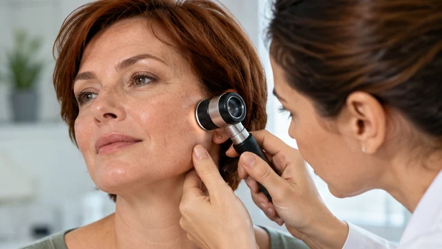
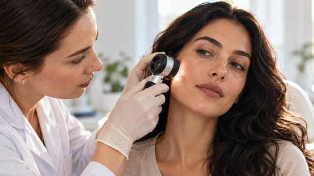
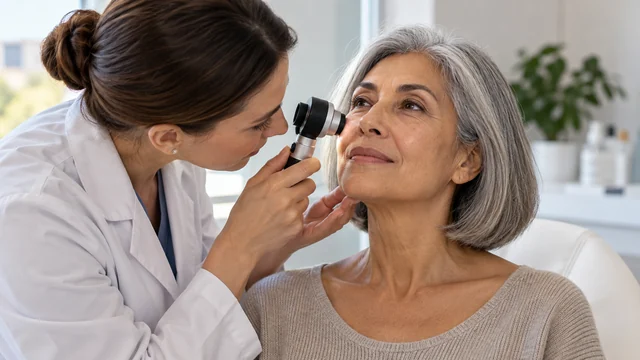
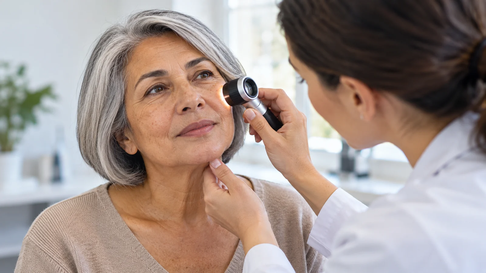
Not every brown spot is a melanocytic naevus. Seborrhoeic keratosis, a vascular lesion, post-inflammatory pigmentation and, less commonly, a malignancy can look similar. Being raised does not prove a spot is harmless. A dermatologist assesses its history, surface and colour and may use a dermatoscope, a lit magnifying instrument that reveals patterns not visible to the naked eye. A phone photograph alone is not a substitute for this assessment.
Watch for the ABCDE signs: A for asymmetry, B for uneven border, C for several colours, D for diameter often over 6 mm (but a melanoma can be smaller), and E for evolution in size, shape or colour. An “ugly duckling” is a spot clearly different from your others. A new lesion in adulthood, unexplained bleeding, a sore or crust that fails to heal, persistent itching or rapid changes also warrant professional attention. ABCDE is a prompt to seek assessment, not a diagnostic test; its absence does not exclude melanoma.
A mole present since childhood that has remained unchanged and comfortable usually does not need urgent removal, although lack of symptoms cannot guarantee safety. Tell your dermatologist if you have a family history of melanoma, many atypical moles, substantial sun damage or immunosuppression: they may suggest an individual surveillance plan, even when the visit is for cosmetic reasons.
What changes the decision: a situation guide
| Your situation | A sensible first step | Why an advertised technique is not enough |
|---|---|---|
| An ordinary stable mole that causes no trouble | Discuss observation or cosmetic removal after examination | The resulting scar may be more visible than the mole |
| A raised mole catches on clothing | Dermatology assessment, then discuss removal | Friction does not establish the diagnosis or required depth of removal |
| A spot grows, changes colour or bleeds without injury | Arrange a prompt in-person examination and discuss biopsy | Destroying tissue without testing may hinder diagnosis |
| Several unusual spots and family risk factors | Have the whole skin examined and agree on surveillance | Removing one mole does not replace monitoring the others |
How is removal chosen, and why is “no stitches” not always better?
The diagnosis, site, depth and purpose determine the method. Surgical excision means removing the lesion with a scalpel and an appropriate amount of tissue; the wound may need stitches. The sample can be sent for histology, which means examination under a microscope. If melanoma is suspected, an adequate, preferably intact specimen is particularly important; the clinician selects the biopsy method and usually favours complete excision where feasible.
A shave removal, in which a protruding lesion is cut across the surface, is sometimes suitable when a clinician judges it benign. Stitches often are not needed, but pigment cells may remain deeper in the skin and the colour can return. This is not automatically suitable for a suspicious flat pigmented spot. Laser treatment, electrodessication or cautery may change some confirmed benign lesions, but destroying tissue can prevent proper histology. “Laser is safer than a scalpel” is not a useful claim without a diagnosis.
Ask about more than the device: what is the likely diagnosis, why is this method suitable, will tissue be available for analysis, and what scar could result? If the diagnosis is uncertain and there is no plan to assess tissue, you have good reason to decline cosmetic destruction. If tissue is submitted, establish who will notify you of the result and what will happen if it is abnormal.
What will the area look like? Healing versus being ready to be seen
For some people the hardest part is a visible dressing on the face, not the procedure. Local anaesthetic injections can sting briefly; you may still feel pressure or touch, but tell the clinician about significant pain. As the anaesthetic wears off, the site may ache, feel tender or swell slightly. Size, body site, technique and your skin all affect the response.
In the first 24 hours, expect the possibility of a little blood spotting, a dressing and redness. With stitches you may see sutures and a cut line; after a shave there is a rounder exposed wound. Do not schedule an important photo session immediately afterward, even if you can resume ordinary tasks the same day.
Over the next days the wound closes gradually; redness, crusting or a dressing may remain visible. The American Academy of Dermatology gives approximately 7–10 days as a guide for many skin-biopsy wounds, but lower-leg wounds and larger procedures can take longer. This does not mean the skin will look unchanged at day ten. Stitches may require a separate visit, while a scar can continue to mature and fade over months.
Once the surface closes, the area may remain pink, paler or darker than neighbouring skin. Mention any history of raised or keloid scars. On the face or décolletage, a fine but visible line may replace a subtle mark. On shoulders, back or chest, skin tension can widen scars. No reliable clinician can promise removal without a trace.
Do not put makeup directly on an open wound unless the clinician approves, or conceal it with aggressive peeling. Care depends on whether there are stitches and dressings. Typical skin-biopsy instructions include gentle cleaning, a thin layer of plain petroleum jelly and a clean dressing; antibiotic ointment is usually unnecessary without a prescription. Ask about immersion in baths, swimming pools and saunas. Once healed, sun protection may reduce long-lasting discoloration. Follow your own clinician's instructions if they differ.
Risks to understand before you consent
Even small removals create a wound. Risks include pain, temporary swelling, bleeding, infection, an adhesive reaction, changes in colour, scarring or regrowth after incomplete removal. Lesions on eyelids, lips, the nose, hands or joints may require special attention to incision placement and function. A pathology result sometimes means more tissue must be removed; that is not necessarily evidence of an error, but a consequence of the diagnosis.
Contact your clinician if pain increases rather than settles, redness spreads, the skin grows hot, pus or marked swelling develops, or you have fever. If bleeding persists after 20 minutes of uninterrupted firm pressure with clean gauze, seek urgent medical assessment; mention any blood-thinning medicines. An atypical or cancerous pathology result requires a definite treatment and follow-up plan, not just scar correction.
An especially important danger is missing cancer when suspicious pigment is destroyed without proper assessment. A before-and-after photo cannot show whether tissue was examined. Diagnosis takes priority over appearance whenever a tumour is suspected. Never cut, tie off, burn or apply purchased acids or online “mole removers” at home: burns, scars, infection and delayed cancer diagnosis can result.
When is observation better, and when should you seek a specialist?
Removal for personal preference may be reasonable for a mole confirmed as benign, but is never compulsory. If the concern is subtle, compare how noticeable the mole is now with the likely mark afterward, particularly on the face where incision lines matter. Trying cosmetic cover can help you judge how much the appearance bothers you, but says nothing about medical safety.
Pregnancy-related hormonal changes can alter some pigmented lesions, but never assume a new or suspicious change is “just hormones.” Discuss prompt examination with a doctor; an elective cosmetic procedure may be rescheduled with your treating clinicians when there is no medical need. Blood thinners, bleeding disorders, diabetes affecting healing, active skin inflammation or an anaesthetic allergy call for an individual plan. Do not stop prescribed medicines yourself.
When comparing centres in Yerevan, look past “five-minute removal”: check the clinician's qualifications, access to examination and dermoscopy, how tissue is sent for histology, how results are communicated, aftercare contacts and an honest description of scarring. Check local licences and credentials through official routes where possible. A device alone does not establish melanoma expertise; a price, address or appointment availability does not prove these safeguards exist.
Seven questions that put you in control
- What did the examination show, and are any features concerning before removal?
- Should my other moles be assessed in view of my history and risk factors?
- Why is excision, shave removal or observation best for this particular spot?
- Will tissue be kept for histology, and who will deliver and explain the result?
- What scar might I have given the site, stitches and my skin?
- When can I realistically wear makeup, swim, exercise and appear in photographs without a dressing?
- Whom should I contact for bleeding, infection, returning pigment or an unexpected biopsy result?
How is this different from removing other skin growths?
With moles, the distinctive decision concerns a melanocytic lesion, its changes over time, and obtaining a reliable tissue diagnosis where cancer is suspected. A soft stalked growth sometimes called a “papilloma,” or a rough age-related plaque, requires a different assessment; see benign skin growth removal. Viral warts and the issue of infection transmission belong under warts and papillomas. One advertised machine cannot safely be selected for every growth before its identity is established.
Cosmetic procedures for tone, skin texture or wrinkles cannot replace examining a suspicious naevus. Sometimes the gentlest choice is a thorough examination, documented photographs and observation without treatment. If a lesion is suspicious, this becomes a medical rather than cosmetic decision and may require a skin-cancer specialist or appropriate surgeon. You can seek another opinion, request a written report and decline an intervention that has not been clearly explained.
Questions readers still ask
Does removing a mole inevitably cause melanoma?
No. Proper removal of a benign mole is not regarded as a cause of melanoma. The danger is mistaking existing cancer for a harmless mole, destroying it without diagnosis or failing to investigate a suspicious lesion.
Should I keep photos of my moles?
Dated photographs under consistent light can help you spot changes between examinations, especially if you have many moles. They cannot establish a diagnosis or replace dermoscopy.
Can brown pigment reappear after removal?
Yes. Pigment-producing cells left behind after a superficial shave can sometimes produce colour again. Show it to a dermatologist, particularly if colour extends beyond the scar or changes; do not try to remove it again at home.