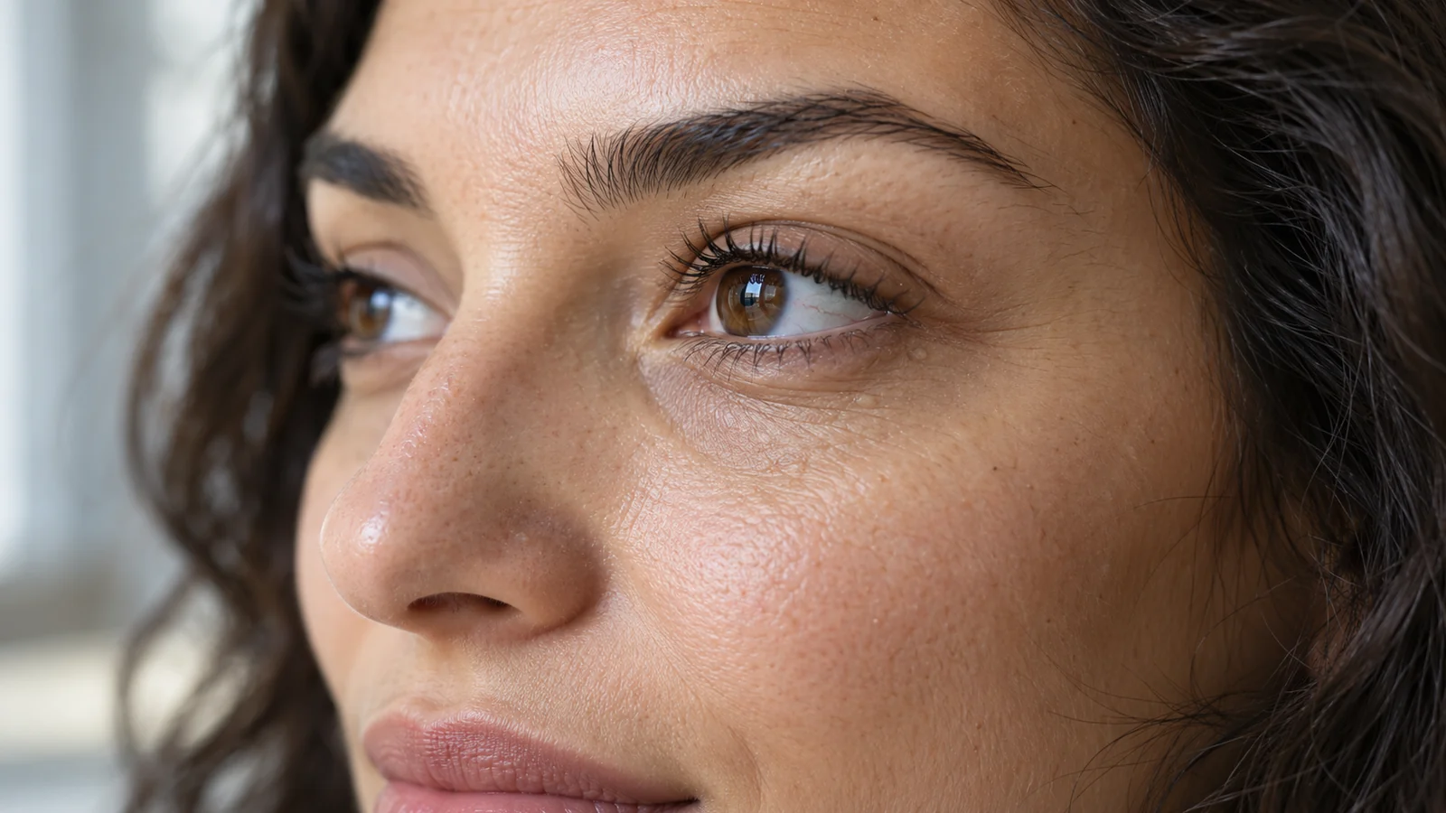

Milia Under the Eyes: Treatment, Skincare or Simply Watching
You notice little white dots under your eyes. Concealer does not disguise their texture, and trying to squeeze them “like spots” achieves nothing. If they are milia—small superficial cysts containing keratin—they are benign and usually need no treatment. Some disappear without intervention. If their appearance bothers you, a dermatologist can examine them and remove suitable lesions with sterile instruments. Near the eyelids, it is safer not to pierce them at home or experiment with acids: thin skin and the nearby eye make mistakes especially costly.
The useful question is not “what should I burn this white dot off with?” but “is it actually a milium, and what is the lowest-risk option this close to the eye?” Similar-looking bumps may need observation, a minor procedure or investigation of another condition. Neither an online picture nor a cosmetic product chosen without diagnosis can answer that question.
Could these be milia? What clues matter?
A typical milium looks like a tiny, firm, whitish or pearl-coloured grain beneath a thin layer of skin, without the open exit of a common blackhead. It is a cyst filled with keratin, a protein in the outer layers of skin—not a fatty lump or a plug of oil you can simply squeeze out. Milia can occur singly or in groups on cheeks, eyelids and under the eyes. Most are neither itchy nor painful.
However, similar pale bumps may be syringomas—benign growths arising from sweat ducts, commonly clustered on lower eyelids. A flat yellowish plaque may be xanthelasma, while swelling near the eyelid edge may be a disorder of eyelid glands. A message or a distant photograph cannot reliably distinguish them. Self-diagnosis is particularly unreliable when bumps are uneven, changing shape or appearing rapidly in groups.
| What you see | What it might be | Sensible next step |
|---|---|---|
| One tiny, firm white spot without pain | Possibly a milium, but examination confirms the diagnosis | Observe it or discuss individual removal with a dermatologist |
| Many nearly identical skin-coloured bumps on lower eyelids | Possible syringomas; extracting “contents” will not solve the problem | Establish the diagnosis before damaging or removing skin |
| Flat yellowish lesions near the inner corner of the eye | Possible xanthelasma, rather than a typical milium | Dermatology evaluation; if confirmed, the clinician can decide whether lipid testing is relevant |
| Growth, bleeding, an ulcer, repeated crusting or loss of eyelashes | Not typical of a simple milium; other eyelid diseases, including rare tumours, need exclusion | Timely in-person examination rather than blind cosmetic removal |
| Red, painful eyelid, pronounced swelling or a vision change | Possible inflammatory or eye condition | Medical assessment, without home puncturing; urgently if vision is affected |
Milia may develop after irritation, injury, burns, rashes or certain topical treatments; these are called secondary milia. Tell the doctor if you have recently used a heavy occlusive cream or had an episode of irritation. The ingredient list of your cosmetics cannot by itself prove what caused one specific bump. Conversely, milia can occur even with careful skin care.
Should you remove them, change skin care or simply watch?
If a pale grain is not growing or inflamed and a clinician finds nothing suspicious, observing it is not a missed opportunity. Milia often clear without treatment, although individual lesions in adults can persist for a long time. “It might disappear” is not the same as “it will go away next week”; the timing depends on the lesion type, skin and other factors. You need not do anything at all if it does not bother you.
If you want a few confirmed milia near an eye removed, a clinician chooses a method based on depth, location and your skin. One direct option is professionally opening the cyst's surface with a sterile instrument and removing the contents. This is not equivalent to squeezing with fingernails or using a cosmetic needle at home: precise identification of the lesion and control close to the eyelid matter. Mention any history of pronounced scarring, pigmentation following inflammation or poor wound healing.
| Approach | When it may make sense | Special limitation under the eyes |
|---|---|---|
| Observation and gentle skin care | A small confirmed milium without warning signs | No guaranteed disappearance or date |
| Targeted professional removal | One or a few confirmed superficial cysts that you want removed | Correct diagnosis, sterile technique, eye protection and scarring risk assessment are needed |
| Clinician-directed topical retinoid | Sometimes considered for numerous milia on suitable skin | May irritate eyelids; not for unsupervised application right beside the eye, and not needed for everyone |
| Device-based or destructive methods | Selected complex or widespread cases after diagnosis | Can damage skin, change pigmentation or harm the eye area; not automatically first choice |
For a single tiny dot, do not automatically agree to aggressive resurfacing, laser treatment or a deep peel of the whole under-eye area. Dermatology references mainly discuss these methods in particular settings, such as very extensive milia. Reviews checked in October 2026 do not present convincing comparative trials identifying one best method specifically for lower-eyelid skin. The potential harm could outweigh the cosmetic benefit.
Why home squeezing is particularly risky on the eyelid
A white tip looks deceptively easy to lift out. In reality, a milium does not have the accessible duct of a familiar acne lesion. Pressure can injure delicate skin and leave a crust, inflammation, a tiny scar or a darker mark. Handling a sharp object only millimetres from the eye also risks injuring the eye itself. An online needle-puncture video is not a safe home instruction.
Harsh scrubs, “acid patches”, vinegar, alcohol and homemade mixtures do not address the depth of the cyst. They can irritate and peel the skin, and getting them in the eye is more than a cosmetic problem. If gentle care has not removed the bump, escalating concentrations without a doctor is not the answer. Do not use wart treatments or other DIY skin-lesion removers on the eyelids either: they are not designed for this use.
Gentle cleansing, avoiding friction and moderate moisturising help keep skin comfortable but have not been proven to dissolve every existing cyst. If a rich cream clearly irritates your skin or feels excessively occlusive, discuss a lighter texture. Reviewing your routine is reasonable, but you have no reason to blame yourself for having used the “wrong” cosmetics.
Will retinol under the eyes help?
“Retinoid” is a broad term for substances related to vitamin A, including adapalene and tretinoin. Some dermatology references describe use for certain cases of widespread milia. This does not mean that any over-the-counter retinol cream will remove a formed cyst on the lower eyelid. Clinical evidence specific to that narrow area is limited; reviews about acne treatment cannot promise a result here.
Eyelid skin irritates easily. Retinoids can cause dryness, redness, stinging and peeling, particularly when applied close to the eye. Do not apply them to the mobile eyelid, lash line or mucous membrane without clear guidance from a clinician familiar with the exact product. A stronger formula does not mean faster or safer treatment. If you are pregnant or planning pregnancy, avoid retinoids for cosmetic skin care; while breastfeeding, discuss medicine choices with your clinician as well. There is no need to start them on your own for one or two spots.
Sometimes someone keeps applying active products, the skin becomes irritated, yet the bump remains. That is a reason to revisit the diagnosis and plan rather than add acids and mechanical extraction. The dermatologist can explain why individual removal or simple observation may better suit your aim at present.
What will the area look like after professional removal?
Social recovery is different from medical recovery. After removal of one superficial milium, a tiny mark, redness or crust may remain visible, for a variable period. Treating several bumps or using a more invasive method can leave more noticeable signs. Do not expect the lower eyelid to look perfectly smooth by the evening of an important photo shoot. Ask the clinician about the likely visibility with your proposed method, without asking for a guarantee.
Sensation depends on the procedure and number of spots: brief discomfort is possible, and the clinician may sometimes choose pain relief. Do not peel off a crust, rub the lid or conceal an unhealed surface against instructions. Ask when you can safely wear makeup and contact lenses, resume skin care and whether a follow-up is needed. If pain, redness or discharge grows after removal, contact a medical professional rather than treating it yourself.
Pigment changes after inflammation can be particularly noticeable in skin prone to post-inflammatory hyperpigmentation. Choice of technique therefore depends on skin phototype, previous inflammatory marks, scars and the depth of the lesion. A gentler plan may offer a better balance of benefit and risk even without a promise of instantly flawless skin.
When is it a medical visit rather than a beauty treatment?
See a dermatologist if a bump is new and unlike earlier ones, is growing, bleeding, repeatedly crusting, changing colour, slow to heal or accompanied by loss of eyelashes. These features do not automatically mean a dangerous disease, but concealing them cosmetically can delay diagnosis. A bump right on the lash margin or inner lid, or one with watering and eye irritation, may call for an ophthalmologist.
Severe eye pain, a sudden change in vision, rapidly developing swelling around the eye or marked inflammation require urgent medical care. That is not the usual course of an uncomplicated milium. Do not expect a white-spot product to treat those symptoms.
For numerous spots that appeared suddenly, lie on a red raised plaque or followed another skin condition, the clinician should assess their type. Some are secondary milia or a rare variant with a different management approach. Mention previous burns, cosmetic procedures, medication and rashes: context helps prevent inappropriate treatment.
How to prepare for a calm consultation
Take a daylight photograph for your own records, but do not diagnose yourself from a zoomed selfie: macro cameras exaggerate texture. Note how long the spots have been present, whether they grow, itch or hurt, and whether they followed a new product or dermatological treatment. Bring the names of active ingredients in your routine and a medication list; mention pregnancy, breastfeeding and previous skin reactions to procedures.
- Are these definitely milia, or is another diagnosis more likely? Is dermoscopy or another check needed?
- What would happen if I did nothing? Is there any sign that makes waiting unwise?
- Where is the safe treatment boundary for this particular lower eyelid, and how will my eye be protected during treatment?
- Could you treat only the individual spots rather than the whole under-eye zone, and what scarring or pigment changes might follow?
- What might my skin look like the next day, when can I use makeup and what symptoms warrant a call?
- If spots recur, is there underlying irritation or another skin disorder to consider?
When comparing offers, do not choose only on a promise to “remove everything without a trace in one visit”. More useful questions are who makes the diagnosis, how they will work near the eye, what risks they disclose and what they will do if a reaction is unusual. Cost depends on the number and position of lesions, the diagnostic complexity and the method; without verified local information, a meaningful price cannot be quoted. If someone rushes you into device treatment before an examination, you can pause or seek another opinion.
What happens next?
Professional removal can clear an existing milium from that particular spot, but does not guarantee that no new ones will appear. Conversely, leaving a milium untreated does not mean that it will certainly keep growing. Prevention depends on the underlying causes and cannot be reduced to one “correct” cream. Protecting skin from injury, gentle care and timely treatment of inflammation make sense, but absolute prevention cannot be promised.
For more practical detail on approaches used at home and by professionals, see the neighbouring guide to how to get rid of milia under the eyes. This page makes the earlier decision easier: recognising what the lesion might be, distinguishing safe observation from removal and understanding the particular risks of the eyelid region. A careful diagnosis is worth more than another irritating product.
A few more questions
Can a milium “infect” neighbouring skin?
No. Ordinary milia are not contagious. Several bumps appearing together does not mean infection is being passed from one to another.
Can you tell a milium from a whitehead by squeezing out its contents?
Do not test this by squeezing. A milium and a closed comedone have different structures; a dermatologist can distinguish them at examination without injuring the eyelid at home.
Does the same approach apply to white dots around a baby's eyes?
No. Milia are common in newborns and often resolve without treatment. Do not apply adult active products or pierce the spots; ask a paediatrician if the diagnosis is uncertain.
Sources and evidence
- DermNet: Milium — clinical appearance, differential diagnosis and treatment options.
- Cleveland Clinic: Milia — patient information about milia and their benign course.
- DermNet: Eyelid skin problems — other eyelid lesions that can resemble milia.
- DermNet: Topical retinoids — benign syringomas and their distribution.
- American Academy of Dermatology: Pregnancy skin care — irritation and safe use issues with topical retinoids.
- American Academy of Dermatology: Sebaceous carcinoma symptoms — pregnancy skin care and retinoids to avoid.