
Benign skin growths: what they are and how they are removed
You may have noticed a small soft “papilloma” on your neck, a rough patch on your back or a growth rubbing against your bra. Online these are often grouped together and offered the same laser treatment. Yet “benign skin growth” is a category, not one diagnosis. Skin tags (acrochordons, or fibroepithelial polyps), seborrhoeic keratoses, viral warts, cysts, melanocytic moles and other lesions can look similar. A few more serious changes can mimic them and need skin cancer ruled out.
The sensible sequence is diagnosis by a dermatologist, a decision about whether tissue testing is needed, and only then a discussion of whether and how to remove the lesion. A confirmed harmless growth can often be left alone if it does not bother you. Removing it may relieve rubbing or change a visible bump, but scarring, pigment change and later development of other similar lesions remain possible. This page explains choices for different non-pigmented or otherwise confirmed benign growths; it is not a home-removal guide.
Why does the diagnosis matter more than the name of the device?
A soft skin tag usually grows on a thin stalk, often on the neck, underarms, beneath the breasts or where clothes rub. A seborrhoeic keratosis often looks rough, waxy or as though stuck onto the skin, and ranges from pale to dark. A viral wart is caused by certain types of human papillomavirus (HPV), so recurrence and transmission need a separate discussion. An epidermoid cyst has a wall beneath the skin; merely emptying its contents may allow it to return.
In everyday speech “papilloma” may refer to either a soft skin tag or a wart, but they are not the same diagnosis. Some lesions are infectious and others are not; some can be removed superficially while others need complete excision. Pigment changes, bleeding, rapid growth or an unusual appearance may call for evaluation for other disease, including malignancy. For melanocytic lesions see the page on moles and naevi; for HPV-related growths, see warts and papillomas.
A guide to similar-looking growths — not a way to diagnose yourself
| What you see or feel | A possible diagnosis the doctor will check | Why it affects treatment |
|---|---|---|
| Soft movable growth on a thin stalk | Skin tag (acrochordon) | A small procedure may suffice, but lookalikes should be excluded |
| Waxy, rough, “stuck-on” patch | Seborrhoeic keratosis | An atypical lesion may need biopsy, not automatic freezing |
| Firm rough bump, sometimes several | Viral wart | Recurrence and transmission need their own treatment plan |
| Rounded bump under the skin, sometimes inflamed | Epidermoid cyst or another lesion | Removing its capsule may be discussed; inflammation changes timing and technique |
| Rapidly changing nodule, ulcer or spontaneous bleeding | Uncertain or potentially dangerous lesion | In-person examination and possible histology first; cosmetic destruction is unsuitable |
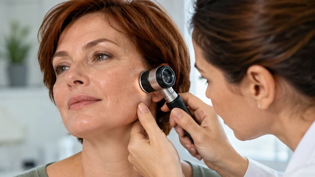
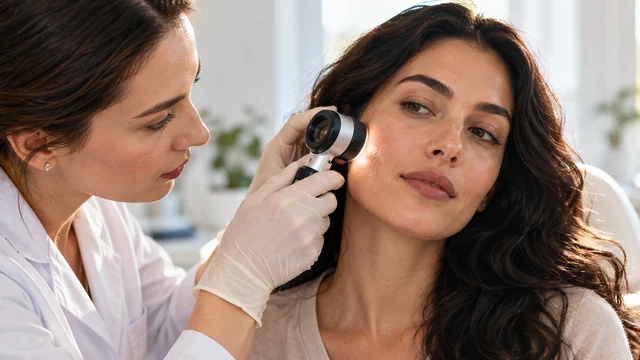
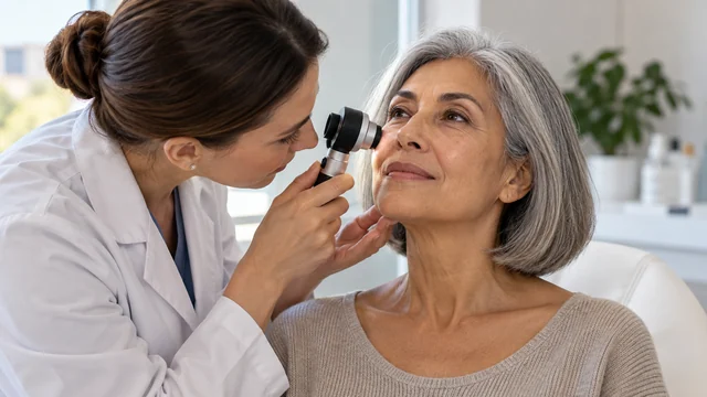
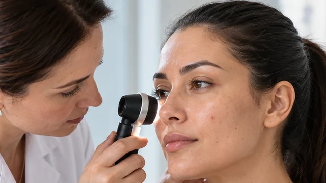
Appearance alone cannot establish a diagnosis. Skin cancers sometimes mimic harmless growths, while benign lesions may bleed simply because they rub against clothing. The rate of change, your medical history, examination of the skin and sometimes dermoscopy or biopsy—a tissue sample examined under a microscope—all matter.
When does removal make sense?
There are usually two reasons: comfort and appearance. A growth may snag on jewellery, a razor or underwear, repeatedly inflame from rubbing or complicate personal care. Or it may be painless but visible, and you want it changed. Either reason is legitimate to discuss. Balance the inconvenience against the possible new mark. A tiny tag hidden in a fold and a broad dark plaque on the décolletage carry different risks of a visible patch afterward.
If a lesion is typical and not troublesome, leaving it alone is often reasonable. Most seborrhoeic keratoses are harmless and require no treatment. Removing one does not prevent a new one elsewhere. Likewise, taking off several skin tags cannot eliminate a person's underlying tendency to develop others, so promises of “permanent removal of all papillomas” are misleading.
Seek diagnosis first for a newly appearing or quickly enlarging lesion, irregular pigmentation, ulceration, bleeding without trauma, a mark unlike your others, increasing pain or failure to heal. Do not wait for it to be “ready for a laser.” Areas near the eye, lips, genitals and mucous membranes need the appropriate specialist's assessment: routine cosmetic methods cannot simply be transferred between sites.
Scissors, freezing, electrosurgery or excision: what is the trade-off?
Once the diagnosis is established, the clinician considers lesion type, base, depth, site and whether tissue needs examination. Cutting off some small skin tags with sterile instruments may involve local anaesthetic and control of bleeding. Cryotherapy means freezing with liquid nitrogen; it is used for some seborrhoeic keratoses and other superficial benign growths. A blister, crust or pale patch may follow.
Electrosurgery and curettage are two ways of treating selected superficial lesions: electrical destruction and careful scraping. A clinician may use them separately or together when appropriate. Surgical excision with stitches may be appropriate for larger or deeper growths, cysts or an uncertain diagnosis. If the identity is unclear, preserving a specimen for histology is a decisive reason to use a tissue-sparing diagnostic technique instead of laser or other destructive treatment. Cosmetic destruction should never replace investigation of possible cancer.
No single technique is best for every so-called papilloma. Freezing may be satisfactory at one site but leave a pale mark at another; excision provides material for diagnosis but can leave a line-shaped scar. A more expensive device does not guarantee a less visible trace. Ask whether only the projecting part or the complete growth will be removed, whether it may recur and what counts as a realistic outcome.
Three decisions to make before treatment
- Diagnosis: exactly what is being removed, and how certain is the clinician without histology? If uncertain, what investigation comes first?
- Skin mark: is avoiding friction more important than a near-invisible surface? Sometimes these goals cannot both be guaranteed by wound healing.
- Extent: one bothersome growth, several similar ones, or first an evaluation of why they appear? Multiple lesions change the plan but do not remove the need to assess each atypical one.
Will it hurt, and when can you appear without a dressing?
Small removals are often performed in an outpatient setting. A local anaesthetic injection may sting briefly; afterward you may sense pressure and touch rather than sharp pain. Freezing may burn or tingle and then leave soreness or a blister. Sensations and the need for anaesthesia depend on the method and site. Tell the clinician if the procedure hurts significantly rather than enduring it to save time.
On the day, the area may look like a pink dot, a small wound, a dressing or a whitened frozen patch. In the following days a crust or blister can develop, and it may be more conspicuous than the original bump. Surface healing commonly takes several days or longer. The AAD gives about 7–10 days for many skin-biopsy wounds, not as a universal deadline for every method, size or body site; folds, friction areas and shins may take longer.
A pink patch, darker or lighter pigment can persist afterward, and deeper removal can leave a scar. Before a wedding, photo session, beach trip or important meeting, allow extra time: being fit to work tomorrow does not mean the site will be invisible. Avoid applying makeup on an open wound, swimming, prolonged heat or friction until your clinician says they are safe. Sun protection after closure may reduce residual pigmentation. Several neck or chest removals can look more obvious socially than one tiny procedure.
Typical skin-biopsy aftercare includes careful washing with mild soap and water, plain petroleum jelly and a protective dressing according to instructions. Do not pick crusts or apply alcohol, acids or antibiotic ointments “just in case.” Rules after freezing or stitched surgery can differ: ask how to care for a blister, change dressings, shower and arrange any follow-up.
What can go wrong, and when should you seek help?
Even when benign, removal may cause bleeding, inflammation, infection, damage to nearby skin, blistering, pigment change, scarring, adhesive or anaesthetic reactions, incomplete removal or recurrence. Risk differs greatly between a thin skin tag, a thick keratosis and a deeper cyst. An inflamed cyst may need a staged or altered plan; never try to lance one yourself.
Increasing pain, spreading redness, heat, pus, fever or bleeding that continues after 20 minutes of uninterrupted firm pressure with clean gauze mean you should promptly contact a clinician. Heavy bleeding or rapid deterioration needs emergency care. If a wound does not heal or a new fast-growing lump appears, do not simply cauterise it again: have the diagnosis reconsidered.
Cutting, tying off, burning or treating an unknown growth with wart remover or online chemicals at home is risky. The US FDA warns that unapproved products advertised for moles and skin lesions can cause injuries, scars and delays in identifying skin cancer. This concerns US regulatory advice, not the registration of individual products in Armenia. A marketing claim that a product is “safe for all papillomas” cannot establish the diagnosis. Wart-removing chemicals can damage healthy skin if applied to a soft skin tag.
When is a procedure not the first choice?
During pregnancy or breastfeeding, an optional cosmetic procedure may be discussed and deferred if appropriate; a suspicious lesion, by contrast, requires timely assessment. Blood thinners, immunosuppressive medicines, clotting disorders, keloid tendency, diabetes with delayed healing and active infection change the plan. Never stop prescribed medicines without the prescribing clinician. Lesions on mucous membranes or in the anogenital area require their own route: genital warts have different rules, and atypical lesions may need biopsy.
If a presumed seborrhoeic keratosis repeatedly bleeds or changes quickly, ask how secure the diagnosis is and why histology is not considered necessary. A second dermatology opinion is reasonable when uncertain. A lesion near the eye may need a clinician familiar with the anatomy. Beauty treatments for glow or texture cannot diagnose an individual skin growth.
Consultation questions that keep the choice yours
- What is the likely diagnosis, and which similar conditions have been ruled out?
- What would happen if we did not remove it now, and how should I monitor it?
- Is histology needed, and who will explain the result?
- Why this technique rather than observation or surgical excision?
- What scar or pigment change is realistic at this site and with my skin type?
- When can I wear tight clothes, swim, exercise and use makeup?
- Does the total price include anaesthesia, histology, dressings, reviews and follow-up? These components matter more than a headline price.
When choosing a clinician, check qualifications, hygiene and facilities, the diagnostic plan and aftercare contact. A photograph taken immediately after removal cannot reveal the scar months later; look for comparable pictures with dates and the actual method. Reviews cannot establish your diagnosis. You can decline removal after the consultation.
Additional questions
Does removing a “papilloma” prevent HPV infection?
A simple soft skin tag is not the same as a viral wart. Even when a wart is confirmed, removing the visible lesion does not guarantee the virus is gone or that no new growth will appear; prevention and treatment depend on the actual diagnosis.
Why send such a tiny lesion for histology?
Small size alone does not prove safety. Histology may be needed when appearance or history does not confidently exclude something more serious. It is not always necessary for every typical skin tag; the clinician decides.
Will a pale ring left by freezing disappear?
It may fade, but lightening can last a long time or be permanent. Discuss this especially if the surrounding skin is darker or the area is highly visible.
Sources
- American Academy of Dermatology: Skin tags.
- American Academy of Dermatology: Seborrheic keratoses — diagnosis and treatment.
- American Academy of Dermatology: Skin biopsy wound care.
- DermNet: Skin tags / acrochordons.
- US FDA: Warnings about misleading dermatology treatments.
- CDC: Anogenital warts — STI treatment guidelines.