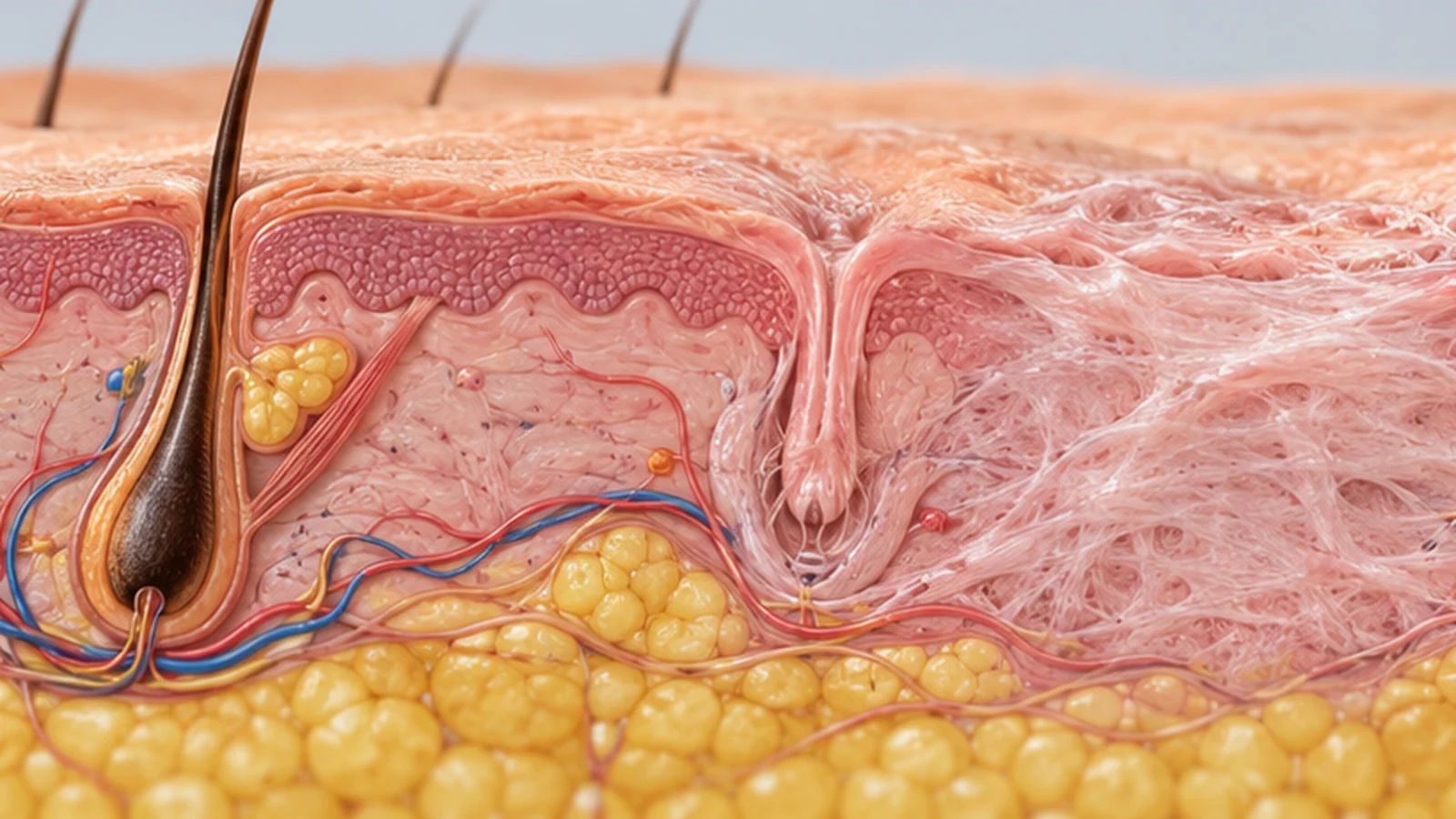

Types of Cicatricial Alopecia: Primary and Secondary Forms
If your dermatologist mentions “scarring alopecia”, it is natural to want to know which form you might have and how it differs from the others. It is not one disease. The first distinction is primary versus secondary scarring alopecia. A widely used working classification of primary forms identifies four categories by the predominant inflammatory cells: lymphocytic, neutrophilic, mixed and nonspecific (unclassifiable). The last means that the sample cannot reliably identify a dominant inflammatory pattern, not that a separate cause has been established. This classification helps clinicians choose investigations and treatment, but you cannot reliably identify a category from the position of a patch or an online picture.
Primary versus secondary: why does it matter for care?
In primary scarring alopecia, the inflammatory process predominantly attacks the hair follicles. In secondary scarring alopecia, follicles are destroyed alongside other skin structures, for example after a deep burn, trauma, radiation, some infections or another skin disease. Either process can leave permanent hairless skin. With a primary type, the immediate question is whether inflammation still threatens surviving follicles. With a secondary type, the underlying cause of skin damage also needs investigation and, where possible, treatment.
“Scarring” describes the result, not necessarily a large visible scar. The scalp may look smooth and pale, or show redness, scale or pustules at the edges. Diffuse thinning or a suddenly appearing smooth round patch can also arise from non-scarring conditions. A subtype therefore depends on the whole pattern, symptoms, trichoscopy and sometimes tissue examination, not a first glance.
The main groups: a quick comparison, not self-diagnosis
| Type or group | Common examples | What the clinician looks for |
|---|---|---|
| Primary lymphocytic | Lichen planopilaris (LPP), frontal fibrosing alopecia (FFA), discoid cutaneous lupus, central centrifugal cicatricial alopecia (CCCA) | Scale and redness around hairs, disappearance of follicular openings, distribution of patches; symptoms can be mild |
| Primary neutrophilic | Folliculitis decalvans, dissecting cellulitis/folliculitis of the scalp | Pustules, crusting, painful nodules, drainage or hair tufts; investigation for infection where indicated |
| Primary mixed | Folliculitis keloidalis of the nape and erosive pustular dermatosis | Different inflammatory cells and features may overlap; further investigation is sometimes necessary |
| Primary nonspecific / unclassifiable | A late-stage or nondiagnostic biopsy in which the predominant inflammatory cell pattern cannot be determined | This does not prove a separate cause; correlate with clinical findings and, where needed, reassess the diagnosis and biopsy site |
| Secondary scarring | Deep trauma, burns, radiation and some infectious, tumour-related or infiltrative processes | Evidence of the underlying injury, medical history, other skin findings and targeted tests |
In this working classification, three categories describe the predominant type of inflammatory cell; the fourth, nonspecific category reflects limits of classifying a particular sample, especially in late “burned-out” disease. Some reviews briefly list only the three identifiable inflammatory patterns. Thus “three inflammatory patterns” and “four classification categories” need not conflict. A biopsy cannot always reveal the original cause, and the cellular picture can change over time.
Lymphocytic types: why can they look so different?
Lichen planopilaris (LPP) can cause irregular patches, itching or scalp tenderness. Redness and tight scale may surround individual hairs. Symptoms can be minimal even while inflammation remains active. The clinician examines the active margin, rather than judging only the size of the already smooth centre. A biopsy may be taken where follicles are still present and inflammation is visible.
Frontal fibrosing alopecia (FFA), regarded as a clinical variant within the LPP spectrum, most often produces gradual recession of the frontal and temporal hairline. In some people eyebrow loss comes first. It is more frequently diagnosed after menopause, but age and hormonal status alone do not establish the diagnosis. Old photographs of your hairline and brows can help document change; classification still requires a dermatologist.
Central centrifugal cicatricial alopecia (CCCA) commonly starts near the crown and expands outwards. It is more frequently reported among women of African ancestry but is not confined to an ethnic group. Hair breakage or itching can be an early clue; in others, thinning is barely noticeable. It is not established that hair styling or chemical products necessarily caused an individual's CCCA; those assumptions are not a diagnosis.
Discoid cutaneous lupus may produce persistent inflamed scaly plaques, colour changes and progressive scarring, sometimes also affecting the face or ears. Having a discoid lupus lesion does not automatically mean a person has systemic lupus erythematosus. A clinician decides whether other investigations are needed based on the rest of the history and examination.
Neutrophilic types: when are there pustules and painful nodules?
Folliculitis decalvans may involve inflamed follicles, pustules and crusts. Several hairs can emerge together as a tuft from one opening. Staphylococcus aureus may contribute in some cases, but the condition is not simply an ordinary contagious infection cured by disinfecting the scalp once. Treatment depends on the clinical picture; swabbing discharge or bacterial culture may be useful.
Dissecting cellulitis/folliculitis of the scalp often causes deeper inflamed nodules and sometimes painful draining cavities followed by scarring. It is not just stubborn dandruff or a pimple under the hair. Squeezing or piercing nodules at home can injure the skin further; worsening pain, spreading inflammation or feeling unwell warrants prompt face-to-face medical assessment.
Some disorders with scars, crusts and inflammatory bumps are placed in the mixed group, such as folliculitis keloidalis of the nape. Appearance does not always correspond to a single inflammatory cell pattern in a biopsy. A category name guides the specialist; it cannot by itself determine whether you need an antibiotic, an anti-inflammatory medicine or an injection.
How are similar-looking patches distinguished in practice?
- Look at the distribution: frontal hairline and eyebrows, crown, scattered patches, nape or sites of prior injury. These are clues, not a definitive test.
- Examine the active margin: remaining hairs, scale around their bases, redness and pustules. This region often gives more useful evidence than an old, completely smooth centre.
- Use trichoscopy: magnification reveals follicular openings, vessels and inflammatory changes. If uncertainty remains, discuss a biopsy and targeted additional investigations.
- Check other possible diagnoses: fungal infection, non-scarring alopecia areata, traction alopecia and female-pattern thinning. More than one condition may coexist.
A small scalp biopsy is done under local anaesthetic but is still a medical procedure that may leave a small scar. Sampling a completely inactive centre can make the original subtype harder to identify. Ask which diagnosis is being tested, where tissue will be collected and what happens if the laboratory description is inconclusive.
Does the subtype change prognosis and daily plans?
Yes, though not in the form of an exact timetable. If inflammation is active, the aim of treatment is to stop or slow damage to still-living follicles and reduce itch, tenderness and new patches. Established scarred areas generally will not regrow hair because of a shampoo or supplement. Some surviving follicles may recover early on, but this is not a guaranteed result.
These diagnoses do not generally require time off work for “recovery from a procedure”. Nonetheless, scalp pain or itching, applying medication and feeling exposed by a widening part or thinning eyebrows can affect daily life. Gentle concealment, a wig or hairpiece that does not pull or press on inflamed skin is an option. Active areas need gentler care; ask about dyeing, tightly pulled styles and cosmetic procedures before proceeding. Hiding a patch visually does not halt the disease.
Hair transplantation is not a universal solution. While inflammation continues, transplanted follicles can also be damaged. Surgery should be considered only after specialist assessment of sustained disease stability and why scarring occurred. Choices among camouflage, observation, anti-inflammatory treatment and potential reconstruction depend on the diagnosis, the area affected and your preferences, not an attractive photograph of someone else's result.
When should you not wait for a routine appointment?
If a patch expands quickly, eyebrows begin thinning, or new pain, burning, redness or pustules appear, contact a dermatologist promptly. Fever, rapidly spreading redness, severe pain, major swelling or general deterioration requires urgent medical help. Progression can occur with little discomfort, so slowly changing patches also deserve examination.
Bring a medication list, details of skin conditions and procedures, and dated photographs showing changes in the hairline. Ask which subtype is suspected and why, whether a biopsy is necessary, how activity will be monitored, what counts as a response to treatment and what to do about new patches. While the diagnosis remains uncertain, it is reasonable to avoid speculative cosmetic procedures. Why scarring alopecia develops is covered separately; this classification is here to support accurate diagnosis, not self-prescribing.
Further questions
Could a diagnosis change after a repeat biopsy?
Yes. Different disorders can share features and inflammation changes over time. If the first biopsy and the clinical picture disagree, the specialist may reconsider medicines, sampling location or the need for additional investigations.
Do neutrophils in a biopsy necessarily mean a contagious bacterial infection?
No. “Neutrophils” names a kind of inflammatory cell, not a confirmed infectious agent. Cultures or other tests are ordered according to findings; treatment is chosen for the disease, not one word in a laboratory report.
Can scarring alopecia exist without an obvious raised scar?
Yes. On the scalp it often appears as missing follicular openings and smooth areas rather than a raised mark. Examination and sometimes further tests are needed for confirmation.
Sources
- JDDG/PubMed — Cicatricial Alopecia Classification
- Dermatol Ther/PubMed — Classification and Histopathology
- DermNet — Folliculitis Decalvans
- DermNet — Dissecting Folliculitis
- AAD — FFA Diagnosis and Treatment
- DermNet — Discoid Lupus
- JAAD/PubMed — The Role of the Scalp Biopsy in the Evaluation of Alopecia
- PMC — Primary Cicatricial Alopecia: Diagnosis and Treatment
- Clinical study (2023) — four working categories of primary scarring alopecia